Diagram Of Hip.and Back.muscles - Reasons Your Hips May Hurt : The hip muscles are all the muscles that act on the hip joint.. Other muscles are small and cover much less space. The bones of the spine and the ribs provide further protection. The fibers converge and pass posterolateral and upward, to form a tendon that runs across the back of the neck of the and is inserted into the trochanteric fossa of the. This is a table of skeletal muscles of the human anatomy. The hip muscles are all the muscles that act on the hip joint.
It is opposite from the chest, and the vertebral column runs down. Hip flexor muscles and attachments. Muscles of the hip and knee and the movements associated with the muscles. Most modern anatomists define 17 of these muscles, although some additional muscles may sometimes be considered. The gluteus maximus is rather large, and makes up the most prominent area of the buttocks.
Hip Muscle Strains Info Florida Orthopaedic Institute from www.floridaortho.com There are anterior muscles diagrams and posterior muscles diagrams. The bones of the spine and the ribs provide further protection. Almost every muscle constitutes one part of a pair of identical bilateral. Most modern anatomists define 17 of these muscles, although some additional muscles may sometimes be considered. It is opposite from the chest, and the vertebral column runs down. • posterior • piriformis • gemellus superior • obturator internus • gemellus inferior • quadratus femoris. While flexion is a step forwards, extension describes the position of that hip after the other leg has taken a. The achilles tendon attaches the muscles of the.
Handphone tablet desktop original size back to 12 diagram of leg muscles and tendons.
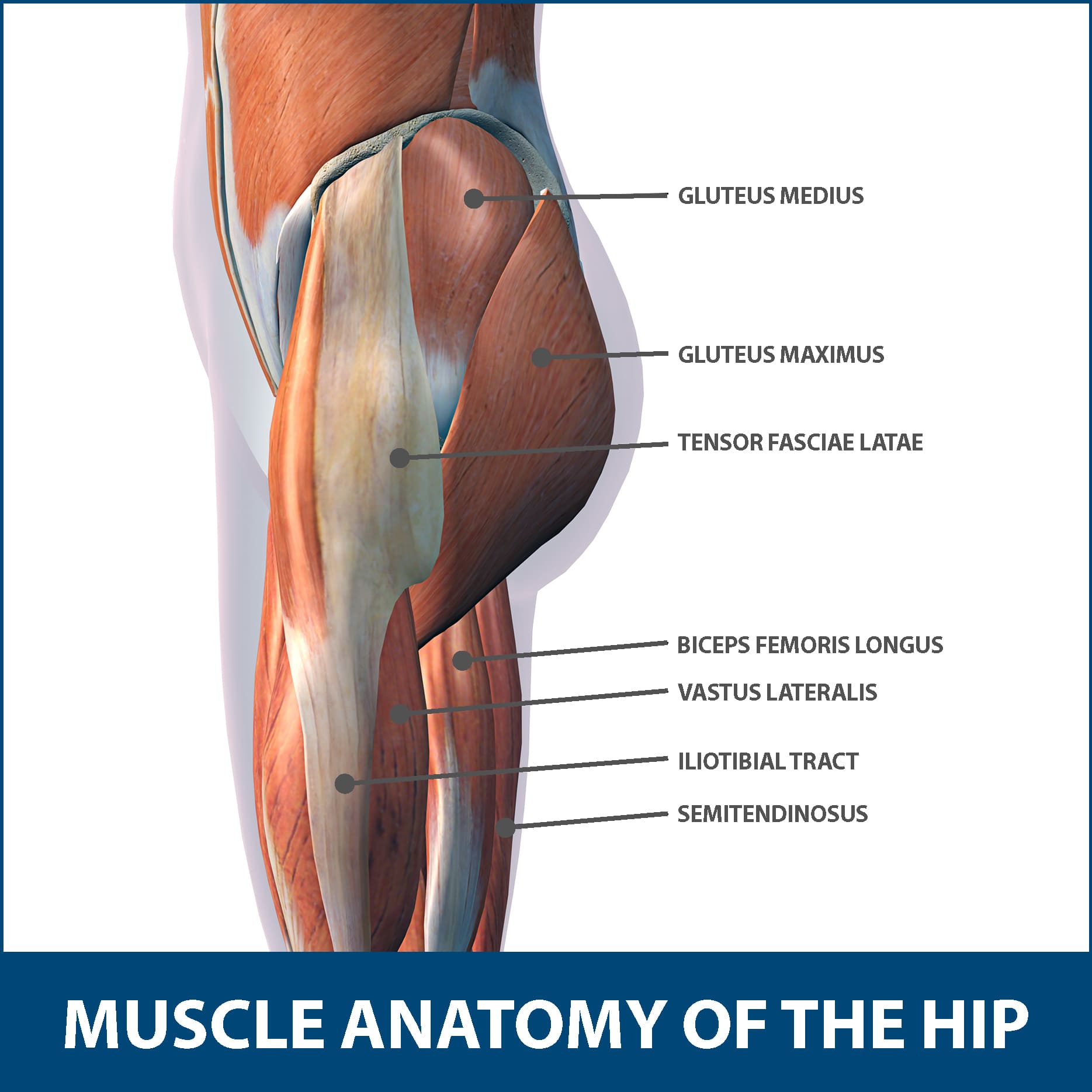
The veins of the upper portion of the back. Common hip and back pain causes include injury to muscles from overuse disc injurydegeneration or spinal stenosis. Back muscles anatomy lower back muscles anatomy human anatomy. Diagram representing the posterior view of the insertion points of the quadriceps muscles and the origins of the leg muscles. • posterior • piriformis • gemellus superior • obturator internus • gemellus inferior • quadratus femoris. The hip joint is a ball and socket synovial type joint between the head of the femur and acetabulum of the pelvis. Nine may seem like quite a lot, but these muscles are essential for creating the wide range of hip movements used by dancers, sportspeople and music lovers. Related posts of muscles of the lower back and hip diagram muscle anatomy posterior. Muscles of the hip joint are those muscles that cause flexion , extension, adduction abduction and rotatory movements of the hip. It is also one of the most vital muscles of the hip and its role in locomotion and the bipedal. Required to throw a baseball, swing a bat or golf club. Insertion because that just makes it more confusing and your muscles don't really identify themselves that way anyhow… These muscles form the pelvic diaphragm which supports and maintains the position of the iliotibial tract and femur.
Other muscles are small and cover much less space. The hip muscles are all the muscles that act on the hip joint. Most modern anatomists define 17 of these muscles, although some additional muscles may sometimes be considered. Globular end of the femoral neck. Here we will look at the gluteal muscles and the inner hip muscles.
Chronic Hip And Back Pain In Hypermobile Dancers The Ballet Blog from www.theballetblog.com Handphone tablet desktop original size back to 12 diagram of leg muscles and tendons. They are attached to the femur (thighbone), tibia (shinbone), and fibula (calf bone) by fibrous tissues called ligaments. The hip muscles are all the muscles that act on the hip joint. Abduction and medial rotation at the hip. It is opposite from the chest, and the vertebral column runs down. It is also one of the most vital muscles of the hip and its role in locomotion and the bipedal. Globular end of the femoral neck. The achilles tendon attaches the muscles of the.
Diagram of muscles and anatomy charts.
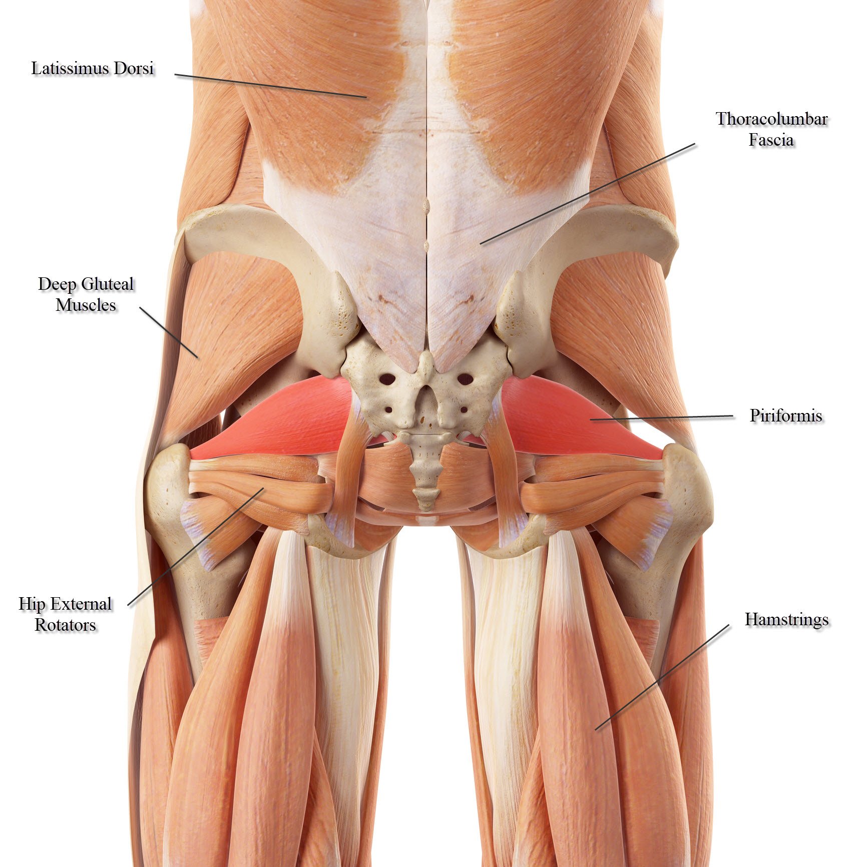
The achilles tendon attaches the muscles of the. Diagram of muscles and anatomy charts. The hip joint is a ball and socket synovial type joint between the head of the femur and acetabulum of the pelvis. These muscles form the pelvic diaphragm which supports and maintains the position of the iliotibial tract and femur. Common hip and back pain causes include injury to muscles from overuse disc injurydegeneration or spinal stenosis. The hip muscle diagram below shows a number of the muscles we will be discussing in the next sections. Muscles/tendons flashcards from molly m. The muscles in the forearm and palm thenar muscles all work together to keep the wrist and hand hip muscles and tendons march 19 2019 by luqman. • the sciatic nerve passes just inferior to the piriformis therefore a tight piriformis muscle my contribute to compression on the sciatic nerve. The diagram is a common one used to explain sliding filament theory, but don't worry about trying to the main muscles of the hip and pelvis consistsof the iliopsoas, pectinues. Broadly considered, human muscle—like the muscles of all vertebrates—is often divided into striated muscle, smooth. This diagram depicts hip muscles and tendons. Common hip and back pain causes include injury to muscles from overuse, disc injury/degeneration, or spinal stenosis.
The hip muscles are all the muscles that act on the hip joint. Each muscle below has the bones in bold for intermediate learners and the specific bony landmarks for advanced learners. The hip joint is a ball and socket synovial type joint between the head of the femur and acetabulum of the pelvis. Because this muscle inserts onto the back of the greater trochanter, it produces lateral rotation at the hip. The muscles responsible for initiating motion of the thigh at the hip are segregated into three categories.
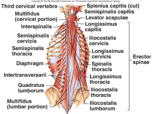
Lower Back Muscle Anatomy And Low Back Pain from ix-cdn.b2e5.com This is a diagram of the larger and more surface muscles of the low back. The muscular system consists of various types of muscle that each play a crucial role in the function of the body. Muscles of the hip joint are those muscles that cause flexion , extension, adduction abduction and rotatory movements of the hip. The bones of the spine and the ribs provide further protection. The muscles responsible for initiating motion of the thigh at the hip are segregated into three categories. The human back extends from the buttocks to the posterior portion of the neck and shoulders. Prime movers cross hip joint anteriorly: The levator ani muscle along with a second muscle forms the pelvic floor.
Here we explain the major skeletal muscles, muscle structure, fibre types, contractions and sliding filament theory.
Most modern anatomists define 17 of these muscles, although some additional muscles may sometimes be considered. Diagram of muscles and anatomy charts. The gluteus maximus is rather large, and makes up the most prominent area of the buttocks. The back comprises the dorsal part of the neck and the torso (dorsal body cavity) from the occipital bone to the top of the tailbone. All of these things can lead to long term back pain (and chronic complaining!). Flexion of hip and vertebral column. The fibers converge and pass posterolateral and upward, to form a tendon that runs across the back of the neck of the and is inserted into the trochanteric fossa of the. • posterior • piriformis • gemellus superior • obturator internus • gemellus inferior • quadratus femoris. Here we will look at the gluteal muscles and the inner hip muscles. Learn with flashcards, games and more — for free. They are attached to the femur (thighbone), tibia (shinbone), and fibula (calf bone) by fibrous tissues called ligaments. Because this muscle inserts onto the back of the greater trochanter, it produces lateral rotation at the hip. This is a table of skeletal muscles of the human anatomy.


0 Komentar